3.2.2 Photoluminescence studies
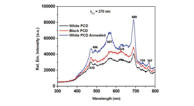
Photoluminescence is a characteristic property of several defect centres in lattice and is thus a confirmatory tool for the presence of defects especially in diamonds. In an attempt to have detailed information regarding non-diamond inclusions, the photoluminescence spectra of all the samples were recorded under excitation of 270 nm wavelength and the spectra is presented in Figure 5.

Figure 5.
Photoluminescence spectra of PCD samples.
The spectra reveal the presence of seven distinct peaks at 470, 496, 567, 625, 689, 734 and 767 nm respectively. Pezzagna et al. [125] suggests that small PL signal for neutral nitrogen vacancy centres is present at 575.67 nm, but for the negatively charged nitrogen vacancy centre, the PL peaks are observed at 637 nm with multiple phonon replica thereafter. It is also well known that silicon vacancy centre emits strong PL signal at 738 nm and a weak replica at 758 nm [121, 126]. Although nitrogen vacancy centres are best photo emitters for diamond but it has strong photon-phonon interactions which is responsible for broad emission spectrum from 600 to 850 nm. That may be the reason for us not getting the nitrogen vacancy signal exactly at the theoretical values in Figure 5. The peaks at the 567, 625 and 689 nm can be attributed to nitrogen vacancy centres, but not definitively. Similarly the peaks at 734 and 767 nm in Figure 5 can be assigned to silicon vacancy centres but again, it is a complex defect centre with an impurity atom and vacancy—so the peaks are not always reproducible at identical positions. Surprisingly there are two more peaks in Figure 5 at 470 and 496 nm which are typically found for Ni-related defect centres—but usually reported for HPHT synthetic diamonds [127]. It is not yet known about the origin of such PL peaks in CVD grown diamonds. Although the presence of the peaks can be identified in all the three samples, variation in the peak intensity indicates the difference in quality of the samples. It is observed that black PCD sample exhibits the stronger peak intensity as compared to that of white PCD. This indicates white PCD contains lesser defect centres and is of better quality as compared to black PCD which is in accordance with the FTIR results. However, upon annealing white PCD sample is seen to exhibit strongest peak as compared to as prepared white PCD and black PCD samples. This signifies that the concentration of defects in white PCD is enhanced upon annealing which may be explained as due to possible aggregation of defect centres. At elevated temperature, the vacancy in a lattice becomes mobile and moves through the crystal and aggregates [128]. Vacancies upon encounter with isolated impurity atoms (nitrogen, silicon), forms the aggregation of defect centres thus resulting in enhanced luminescence.


 Whatsapp/wechat